Today, I’d like to introduce a very special front tooth crown case. This is the story of replacing the front tooth crown for a 54-year-old Korean-Japanese female patient living in Japan. She had visited several dental clinics in Japan hoping to replace the old crown on her upper front tooth, but was reportedly refused because it would be “difficult to do the crown again.”
Then, after accidentally coming across photos of aesthetic prosthetic cases from our clinic, she decided to receive treatment in Korea. Since she lived overseas, it was difficult for her to visit Korea frequently, so a treatment plan had to be made with that in mind. I’ll explain in detail what process was followed and what results were achieved.
Initial examination – panoramic X-ray and facial assessment
When she first visited, we took a panoramic X-ray to check her overall condition. The results showed that the four teeth from the upper lateral incisor to the opposite lateral incisor already had posts and cores placed after root canal treatment. The right canine had only been treated up to the core stage after root canal treatment and was left without a crown, while the left canine had been restored with resin.
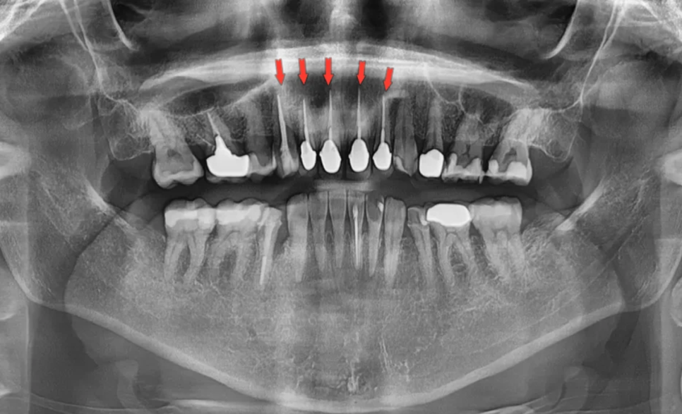
A total of five teeth had undergone root canal treatment
In the facial assessment, several important features were observed. First, she had a deep bite with the front teeth biting down very deeply, and the upper and lower jawbones were developed in a way that made it somewhat difficult to close her lips comfortably. When she smiled, her smile line showed both canines, so for an aesthetically pleasing smile, it seemed appropriate to include the canines in the prosthetic range. In addition, her skin tone was somewhat darker, so this also had to be considered when selecting the crown shade.
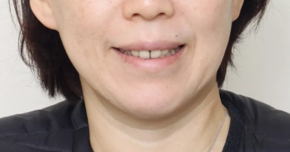
Both canines are visible when smiling
Intraoral examination – problems with the existing crowns and tooth condition
When we examined the inside of the mouth in detail, there were quite a few issues. In summary, they were as follows.
| Problem item | Details |
|---|
| Existing crown condition | Marginal discrepancy, discoloration, and uneven color between teeth in old porcelain-fused-to-metal (PFM) crowns |
| Space between front teeth | Spaces between the two central incisors and between the left front teeth |
| Right canine | Discoloration, wear near the gum line (enamel wear), gingival recession |
| Left canine | Worn incisal edge (tip) |
| Common issue on both canines | Excessive inward tilting of the tooth axis |
| Black triangle | Black triangular space between the right incisor, lateral incisor, and canine |
| Midline | The center lines of the upper and lower teeth were not aligned |
In short, this was a case with many areas that needed aesthetic improvement. The reason she was likely turned down by dental clinics in Japan was probably not only the severe damage to the existing teeth (post + core), but also these complex combined issues.
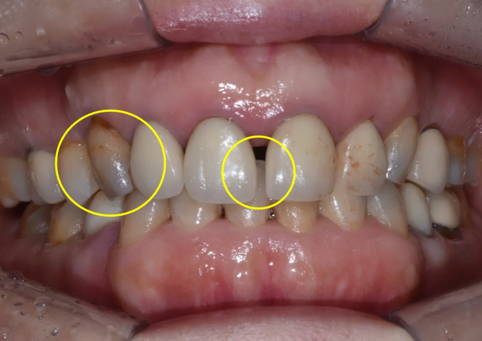
There is a very large space between the front teeth, and the canine on the patient’s right side is severely discolored
The canines are tilted significantly inward
The spaces between the teeth (black triangles) are also quite severe
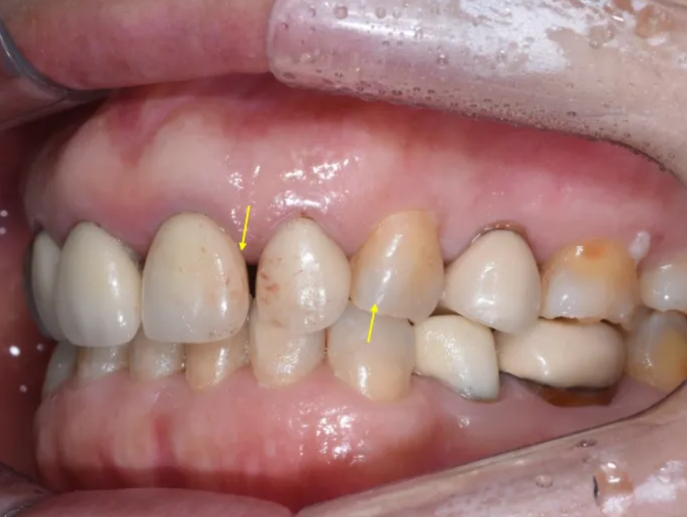
Spaces between the teeth are visible, and the canines are quite worn
Treatment plan – why PFZ crowns were chosen
After putting the examination results together, we made the following treatment plan.
The treatment area covered a total of six teeth, from the right canine to the left canine. Four existing crowns would be replaced, and new crowns would be made for both canines.
For the prosthetic material, we chose PFZ (Porcelain Fused to Zirconia) crowns. PFZ crowns are crowns in which porcelain is layered over a very strong zirconia material. I’ll explain why PFZ was chosen for this patient by comparing it with the existing PFM crowns.
| Category | PFM crown (existing) | PFZ crown (chosen) |
|---|
| Internal framework | Metal | Zirconia |
| Strength | Moderate | Very high |
| Aesthetics | Risk of metal margin exposure and gum discoloration | Natural color without metal |
| Suitable case | General cases | Suitable for patients with deep bite and strong bite force |
Because this patient had a deep bite and strong biting force on the front teeth, the high strength of zirconia was essential. At the same time, since this was the front tooth area, aesthetics were also important. PFZ crowns, which allow porcelain to be carefully layered over zirconia to achieve translucency and color similar to natural teeth, were the best choice.
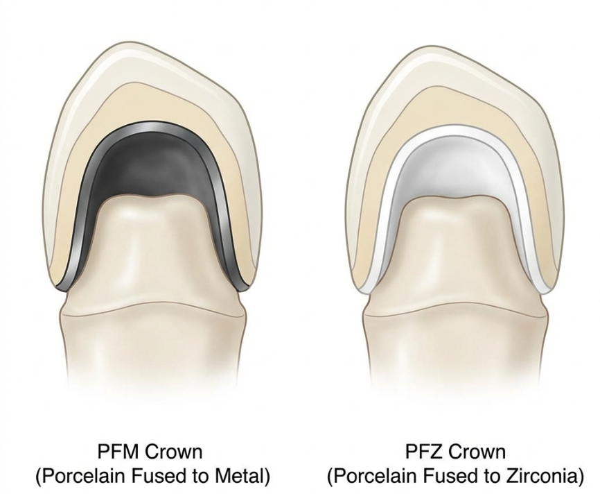
PFZ crown – porcelain is layered over a zirconia coping to achieve both aesthetics and strength
Before starting treatment, we explained the important matters to the patient in advance. We told her that when removing the old crown, the condition of the internal abutment tooth could be worse than expected, and in the worst case, extraction might be necessary. We also informed her that, because she was coming from overseas, she would need to live with temporary crowns for about three months.
Treatment process STEP 1 – Removing the existing crowns and checking the internal condition
First, we carefully removed the old PFM crowns. This process of checking the condition of the tooth under the crown is actually the most nerve-wracking moment.
After removal, we found severe discoloration in the internal tooth, as expected, and secondary caries (new decay that developed under the crown) as well. Fortunately, the post-core had not come off and the tooth root was not fractured, so we were able to continue treatment without extraction.
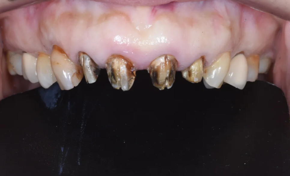
Internal condition after crown removal – severe discoloration and secondary caries are confirmed
As a reference, it is not unusual to find these kinds of internal problems when replacing old crowns. Even if the crown looks fine from the outside, bacteria can enter through tiny gaps between the crown and the tooth, allowing decay to progress quietly from the inside. That is why it is important to have old crowns checked regularly.
Treatment process STEP 2 – Caries removal and core buildup
Next, we thoroughly removed the decay that had been found. After that, we performed a core buildup using light-colored resin.
A core buildup is the process of filling the damaged parts of the tooth with a material such as resin to create a stable post that can support the crown. The reason we used a light-toned resin material was to prevent dark color from showing through the final crown. Especially in the front tooth area, where aesthetics are important, such details have a major impact on the result.
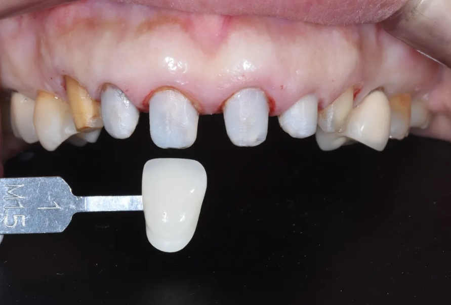
After removing the decay, the core buildup was completed with light-colored resin
Although there was not enough remaining tooth structure, the core buildup provided reinforcement and secured the support needed to keep the crown stable. After that, we shaped the teeth (abutment preparation), took impressions, and fitted temporary crowns. The patient returned to Japan and lived with the temporary crowns for about three months.
The key design points – aesthetic prosthetics for deep bite and alveolar bone prominence
There were several key points we paid special attention to when designing the crowns in this case. Unlike a standard front tooth crown, many aspects required a different approach.
- Considering the prominence of the upper jaw bone
Since this patient had a protruding upper jaw bone, the key was to design the contour of the crowns as flat as possible. If the crowns were too bulky, the lips could appear more protruded. We also paid close attention to angle design so that the tooth axis would not tilt outward.
- Central incisors – splint crown design
There was space between the two central incisors, and to prevent that space from opening again, we designed them as connected crowns.
- Improving black triangles
To reduce the black triangular spaces visible between the teeth, we carefully designed the crown shape to match the interdental spaces.
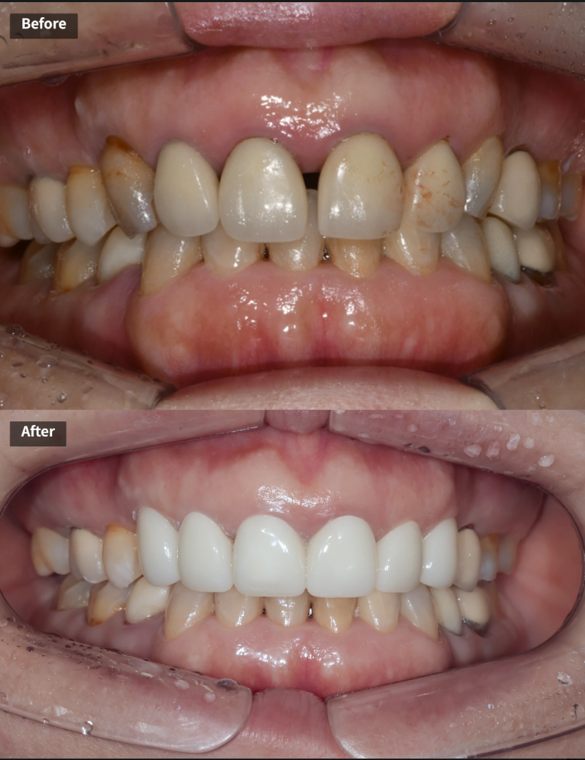
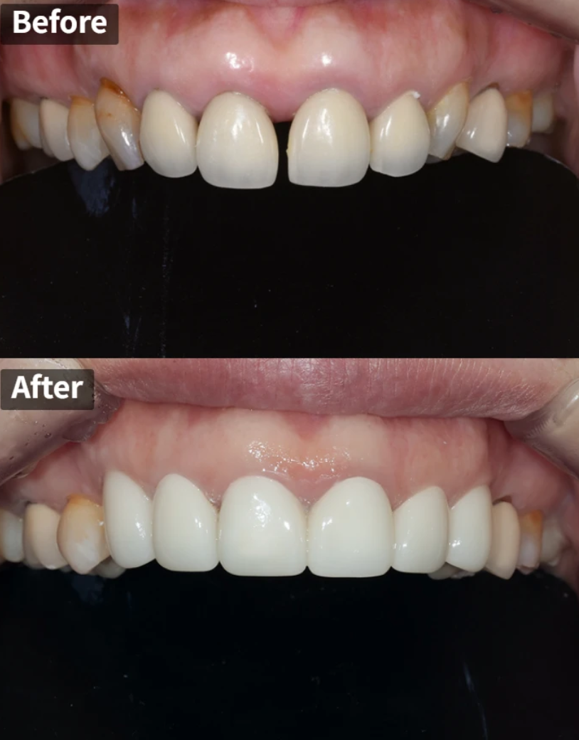
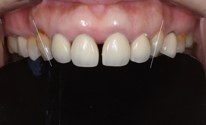
Final result – a natural and harmonious set of front teeth
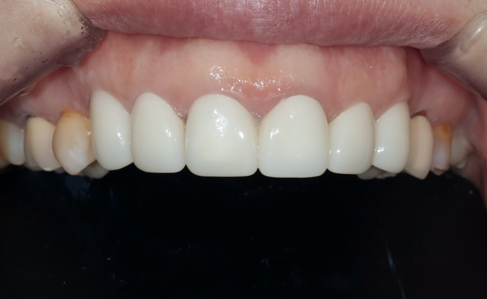
Final prosthetics completed – all the teeth look naturally aligned. The two central incisors were made as connected crowns
After about three months with temporary crowns, the patient visited Korea again, and the final six PFZ crowns were placed.
The comparison before and after treatment showed the following improvements.
| Before treatment | After treatment |
|---|
| Black triangular spaces between teeth (black triangles) | Major improvement in black triangles |
| Gaps between front teeth | Spaces resolved, neat alignment |
| Crown discoloration, uneven color | Natural and uniform shade |
| Crown marginal discrepancy | Precise marginal fit |
| Risk of front teeth separating again | Re-separation prevented with connected crowns |
| Concern about lip protrusion | Improved lip line with flatter crown contours |
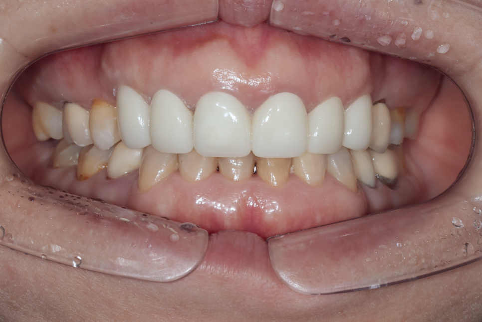
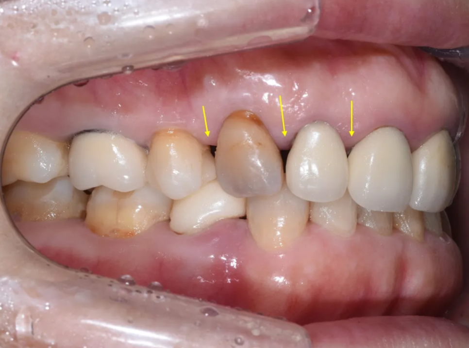
A result that also blends naturally with the lower teeth
Treatment review and what we can learn from this case
Let me summarize a few important points from this case.
First, when removing old crowns, the internal condition may not be good.
When a crown is opened, decay may already be progressing inside, or in severe cases, the tooth post itself may come loose, leading to a situation where extraction and implant treatment become necessary. Therefore, it is important to understand these possibilities thoroughly before starting treatment.
Second, for patients living overseas, coordinating the treatment schedule is key.
As in this patient’s case, there may be situations where they must live with temporary crowns for nearly three months, so it is necessary to understand how to manage temporary crowns carefully.
In this patient’s case, fortunately, the condition of the abutment teeth was not severe enough to require extraction, and they could be well reinforced with a core buildup. Above all, thanks to the patient’s patience and cooperation during the long three-month temporary crown period, we were able to achieve a satisfying result. Although this was a case that had been turned down by several clinics in Japan, it was successfully completed through a systematic treatment plan and precise procedure.
Don’t miss the replacement time for old front tooth crowns
Old porcelain-fused-to-metal (PFM) crowns are at increased risk of marginal discrepancy, discoloration, and secondary caries after 10 to 20 years. Even if everything looks fine on the outside, problems may be progressing quietly inside the crown, so I strongly recommend regular dental checkups.
Today, with modern prosthetic materials such as PFZ crowns that combine aesthetics and durability, even cases that were difficult in the past can achieve very good results.
If you have been told that something is “difficult” elsewhere, please do not give up and consider getting another consultation. There may be a way.
Thank you. This was Dr. Kim Sun-min, Director of Hakdong Station Ceramic Dental Clinic.