Hello,
This is Yonsei Chorokbit Dental Clinic.
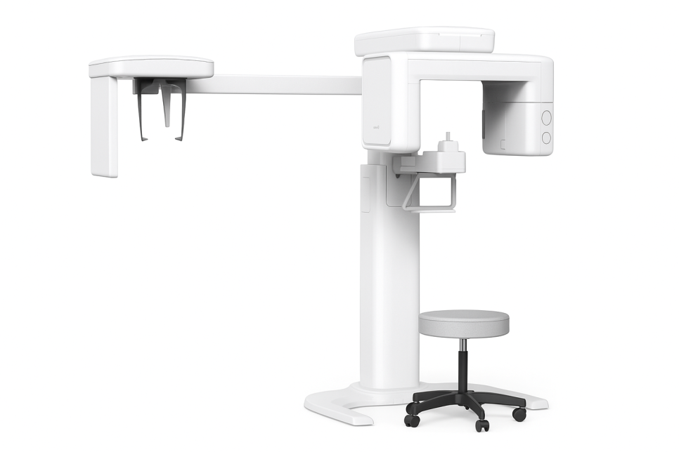
The first step in dental treatment is
X-ray imaging.
Everyone has probably
had an oral X-ray taken at least once.
Have you ever wondered
why it is necessary to take images of areas
that seem fine on the surface?
The inside of our mouths is made up of a much more
complex structure than it may seem.
Because the visible part is only a portion of the overall structure,
the root area and the inside of the gum bone (alveolar bone)
cannot be seen from the outside.
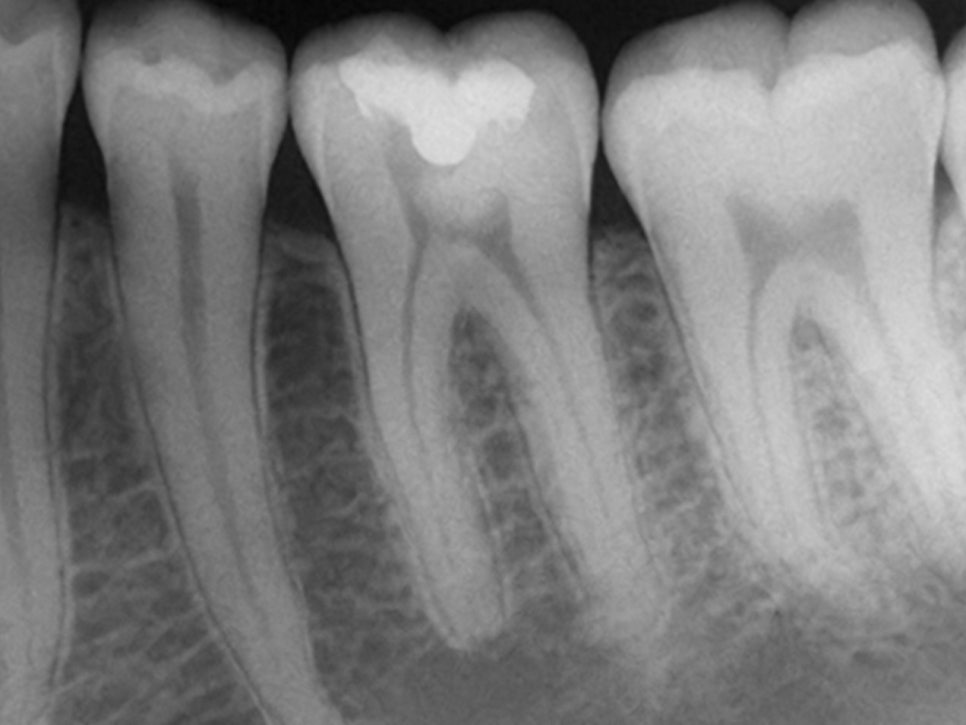
- To detect problems that cannot be seen
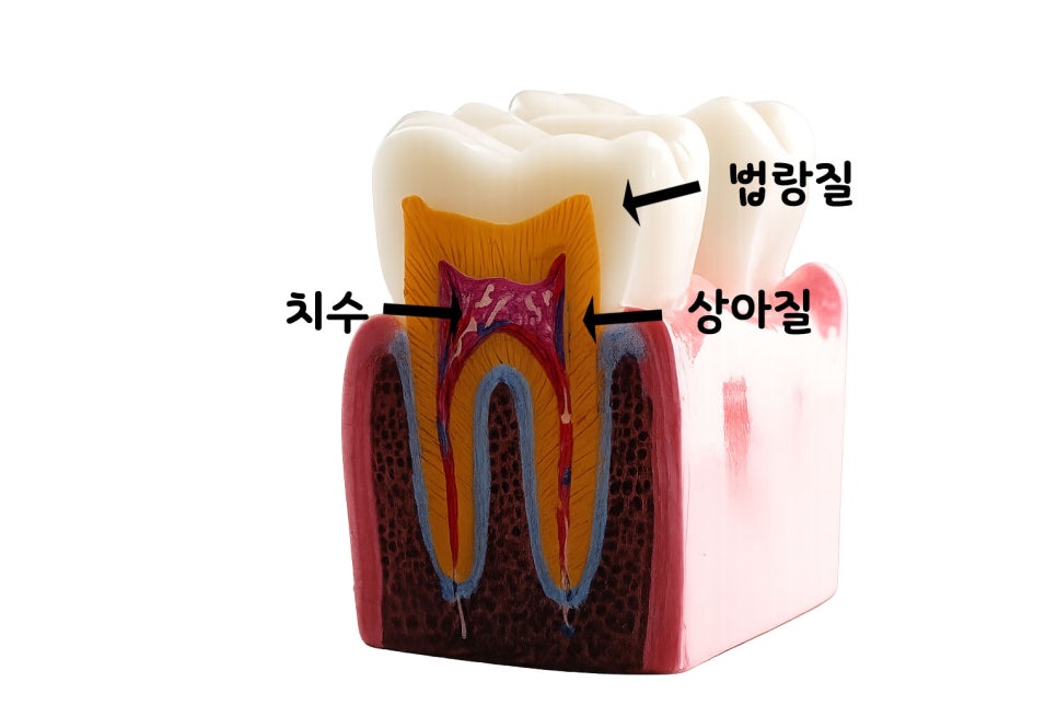
The outer layer of a natural tooth is covered by a hard tissue
called enamel.
If you look at the photo prepared by Yonsei Chorokbit Dental Clinic,
you can see dentin and pulp beneath the enamel,
a nd even if the tooth appears healthy on the outside,
problems may already be progressing in these areas.
In particular, early-stage cavities (caries)
often do not show up at all.
If they start in the side surface (proximal surface) or the deep grooves of the chewing surface (occlusal surface),
they are difficult to detect with the naked eye
until they have progressed deeply.
These hidden cavities can be found early through X-rays,
allowing them to be treated with simple procedures.
In addition, inflammation hidden inside the bone,
or infection at the root tip,
can only be confirmed through X-rays.
In most cases, patients do not know about these lesions
until symptoms appear.
Therefore, it is most important to receive regular checkups periodically.
- Understanding the teeth and surrounding tissues
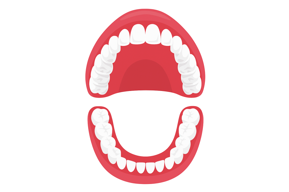
In our mouth there are
32 permanent teeth, including the wisdom teeth,
each with a different shape and size.
To briefly explain it, as prepared by Yonsei Chorokbit Dental Clinic,
the front teeth play the role of cutting food,
the canines tear food,
and the molars grind and crush food.
For them to function properly,
the roots must be firmly anchored in the bone.
At this point, X-rays can be used to evaluate the root length, shape,
and stability,
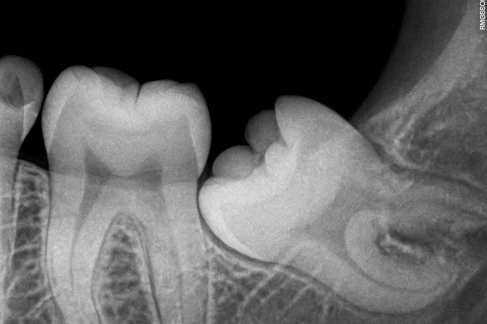
and when teeth have not yet erupted, such as impacted wisdom teeth,
their direction and position can also be checked,
making it possible to predict and prepare for potential future problems in advance.
- Establishing a safe and accurate treatment plan
Treatment must be carried out precisely, and especially before implant placement or tooth extraction,
it is important to identify the exact anatomical structures in advance.
For example, near the lower molars there is a canal through which a nerve passes.
If treatment is performed without knowing the exact location of this structure,
complications may occur.
Therefore, through X-rays, it is possible to know the location of such structures in advance
and establish a safe treatment plan.
Yonsei Chorokbit Dental Clinic also uses them to evaluate whether inflammation is healing well after treatment,
and whether the implant is bonding well with the bone.
- Types of imaging and their characteristics
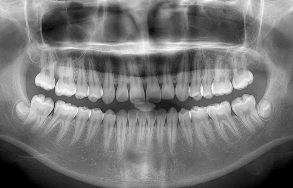
When you want to see the entire mouth at a glance,
a panoramic X-ray is taken.
This is taken by rotating around the head, allowing the condition of all teeth and the jawbone to be checked,
and the position of wisdom teeth, the temporomandibular joint, and sinus conditions
can also be observed together.
However, because there may be distortion,
when you want to look closely at a single tooth,
small films are bitten between the teeth to take images of only 2–3 teeth,
allowing the condition of the relevant area and surrounding tissues to be examined.
- Is it safe?
Many people worry about radiation exposure,
but the amount used here is much lower than the radiation
we are naturally exposed to in daily life.

According to the materials prepared by Yonsei Chorokbit Dental Clinic,
both panoramic X-rays and CT scans are at levels lower than cosmic radiation received on a round trip to Europe.
In addition, since the imaging time has become shorter than before,
it is now possible to obtain clearer images while minimizing the burden on patients.
Pregnant women and children require caution, but imaging is performed only when absolutely necessary
and in the minimum amount needed,
and exposure to other areas can be blocked by using protective equipment such as a lead apron.
- Why do I need a checkup every 6 months?
Even when there does not appear to be any problem on the surface,
hidden early lesions can be found,
so we recommend having a checkup once every 6 months to 1 year.
In particular, people who have previously experienced cavities
or gum disease, or who smoke,
are at higher risk of developing oral problems,
so it is better to have examinations more often.
The reason for taking X-rays during treatment
is not simply as a routine test.
They are a diagnostic tool used to accurately identify the inside that cannot be seen with the eyes
and the surrounding tissues,
in order to determine the appropriate treatment method.
Therefore, please do not postpone your checkups
and make sure to receive them regularly.
I hope the information I shared today was helpful,
and I will end my explanation here.
This has been Yonsei Chorokbit Dental Clinic.
Thank you.