"Because there is one more nerve,
improvement may be a bit difficult."
.
.
.
Retreatment root canal therapy is known as the last resort for saving a natural tooth.
While the success rate is not low,
due to the tooth’s structure, it often ends in failure.
One of the reasons is the canal called "MB2."
This refers to the presence of two thin canals in the mesiobuccal root of the maxillary first molar.
According to several papers, about 95% of people have an MB2 canal.
In the textbook approach, it is correct to find it clearly and remove the tissue,
but
it can be difficult to detect, and there are cases where calcification makes the opening hard to find.
So it is often left untreated, but usually, treating just one of the two canals in the mesiobuccal root does not cause any major problems.
However, there are times when bacteria or germs enter through an untreated MB2 and become a route of infection.
Then, even after root canal treatment, throbbing pain can occur, causing discomfort.
In such cases, even if the opening is difficult to find and the canal is calcified,
finding the MB2 becomes a key point that determines success or failure.
In a case with such a complex structure,
I will share related information from Dangsan-dong Dental Clinic on how to find the opening and lead the treatment to success.
Point 1: Accurate diagnostic data
To observe the inside of the tooth,
radiographic data is needed.
Based on panoramic and CBCT data,
you can examine the tooth in detail.
Directly creating a pathway to observe the inside would be the most accurate,
but
accurate preoperative assessment using data is necessary to increase the success rate of treatment,
so the step of analyzing based on precise data must come first.
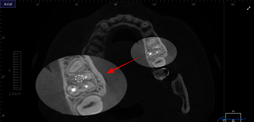
If you look at the CBCT data prepared by Dangsan-dong Dental Clinic,
you will see a round structure between the three canals filled with white material.
This area may be suspected to be the hidden MB2.
Normally, the internal pathway is visible clearly because it is radiolucent,
but here, the pathway inside is not visible at all.
In such cases, calcification can be suspected.
If calcification progresses, even if the opening is found, it can be difficult for instruments to enter,
so the treatment process may become somewhat more complicated and longer.
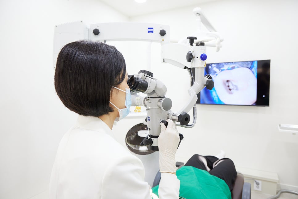
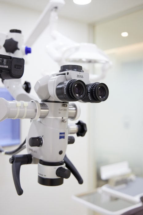
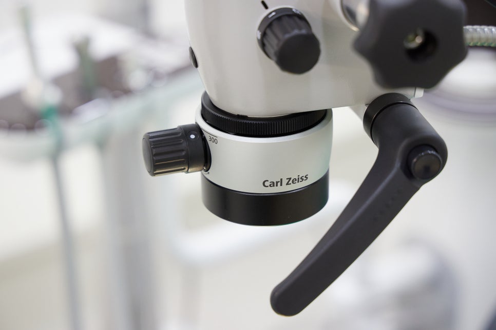
Point 2: Microscopic microscope
After identifying the location of the hidden structure based on preoperative data,
retreatment root canal therapy begins.
Teeth are much smaller than you might think.
The structures inside them are even more delicate.
So in this kind of difficult and delicate process,
a [microscopic microscope] can be used.
It allows for a much more detailed observation of small structures,
which greatly helps in finding very small openings.
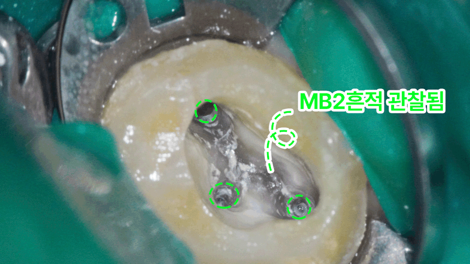
If you look at the data prepared by Dangsan-dong Dental Clinic,
the inside of the molar is shown very large and in great detail.
This is because a microscopic microscope was used.
You can clearly see the three canals in the photo, right?
I can see traces of the MB2 canal between them.
A small circular pathway is visible,
and if you scrape this area with a fine instrument,
you can find the opening.
Because it is very small, using a microscopic microscope is much more advantageous than viewing it with the naked eye when looking for traces.
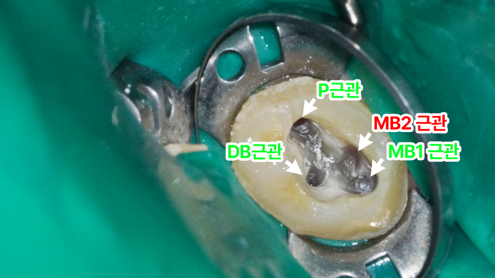
If you look at the area marked by Dangsan-dong Dental Clinic,
further scraping the area with only traces makes the MB2 opening
clearly visible.
Because a microscopic microscope was used,
it was relatively easier to find the trace and reach the opening.
If only the naked eye or loupes had been used,
the process would have been a little more difficult and tiring.
Point 3: MTA restoration
After finding and improving a structure that was difficult to locate,
if the infection is resolved,
the inside is filled with material to achieve sealing.
If there is a gap between the canal wall and the material,
the risk of reinfection later is high,
so achieving a seal during the restoration stage is truly important.
MTA can be considered an appropriate material for this purpose.
I will explain this in detail at Dangsan-dong Dental Clinic.
It has excellent sealing ability and the advantage of being biocompatible,
making it one of the materials actively used in conservative dental treatment.
The biggest reason MTA is preferentially chosen in advanced cases such as apexification in immature permanent teeth,
cases with a wide apical area, or perforations,
is its outstanding sealing ability.
Because of MTA’s advantages and good prognosis,
the American Association of Endodontists (AAE) and the Korean Academy of Endodontics also recommend its use.
Point 4: Use of a rubber dam
Lastly, among isolation methods,
the rubber dam is known to be the most effective.
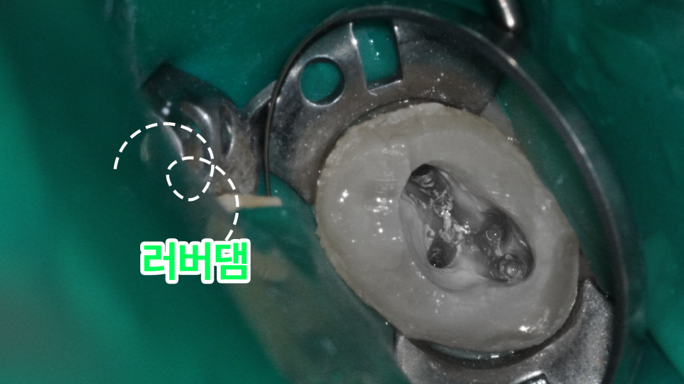
As in the photo from Dangsan-dong Dental Clinic,
it is used to isolate only the area that needs treatment,
and
when moisture control is especially important, as introduced today,
it can be said to be essential.
Without moisture control,
the bacteria present in the mouth are more likely to enter and contaminate the inside,
so it is definitely necessary.
Today, at Dangsan-dong Dental Clinic, we shared information about the MB2 canal, which interferes with the success of retreatment root canal therapy.
There may be limitations due to the tooth’s structure,
but because the value of a natural tooth is greater than anything else,
I believe we must use various methods to overcome those limitations and lead the treatment to success.
I hope this post was helpful, and I will wrap up here.
This has been Kim Min-young. Thank you.