Hello, I’m Kim Min-young, a specialist in restorative dentistry.
Mongolians are said to have excellent eyesight.
So they can see things far away very well.
Conversely, people with poor eyesight
wear glasses or use magnifying glasses
to see distant or small objects better.
Because eyesight has limits,
aid tools are used
to observe things in more detail.
The same is true during treatment.
Molars are smaller than a thumbnail.
In many cases, it is difficult to look closely at
the tiny internal tissues with the naked eye.
That is why, in such cases, an auxiliary tool
called a dental microscope in Dangsan-dong
can be used.
It is especially necessary for cases that require careful attention,
such as molar fractures or root canal treatment.
Very small internal structures of a natural tooth
Based on the lower second molar,
men: 19.0 mm
women: 18.2 mm
This is the average total tooth length.
It is much smaller than you might think, right?
Root canal treatment, which requires looking into and improving
very small structures inside such a small molar,
and fracture cases, where very small cracks or broken areas
inside a natural tooth must be identified,
require careful observation of detailed elements,
so I believe a dental clinic in Dangsan-dong truly needs this.
And this is not just my own opinion.

At the 2012 American Association of Endodontists meeting,
magnifying devices such as microscopes were presented
as an indispensable and important part
of improving the inside of root canals.
Many restorative dentistry specialists also
recognize this.
That is because a problem can only be recognized by looking at it,
and it can be corrected accurately only by examining it in detail.
How is it used in treatment?
I will explain what kind of help it actually provides
at a dental clinic in Dangsan-dong.
A case in which a tooth with a crown
continues to hurt and feel sensitive.
It may not simply be a symptom caused by
the stress applied during treatment,
which made the internal structure sensitive,
but rather a very small defect in the prosthesis itself.
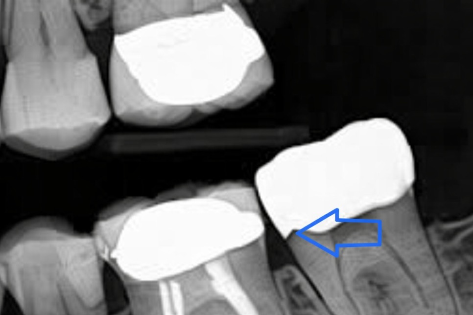
If, as shown in the photo, the transition area between the crown and the tooth
is not tight enough (open margin)
and the pulp becomes congested,
it is necessary to carefully look between the crown and the molar
to find the cause of the problem and improve it.
Because it is such a small issue,
using a microscope during the diagnostic process
is a great help in identifying the cause.
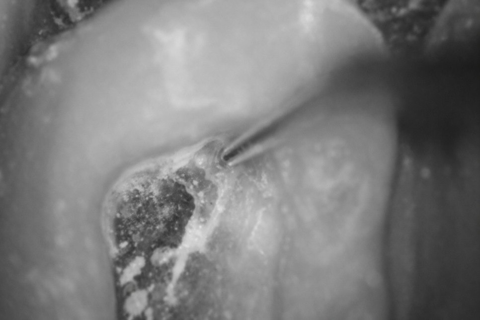
Also, if there is no cavity or inflammation,
and the prosthesis and other elements are normal,
but the pain continues,
there is a possibility that a very small crack has formed.
It helps detect tooth fractures that occur in the transition area
between a previously placed prosthesis and the tooth,
or on the distal side of a molar that is difficult to observe with the naked eye.
It is a great help not only in the diagnostic and observation process
but also during treatment.
It does not simply play a role in finding problems.
Improving problems that are difficult to find is even harder.
This helps the process proceed more smoothly.
Previously, at a dental clinic in Dangsan-dong, I shared information
about MB2, which is one of the factors that interferes with root canal treatment.
🔽Related article URL🔽
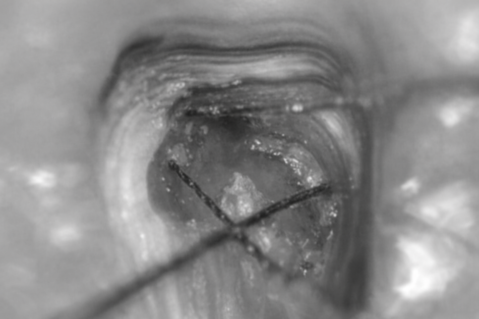
In cases like this, due to structural limitations,
when it is difficult to find the canal orifice,
or when the root canal has already been exposed because of decay,
or when the DL root is far from the others and therefore difficult to find,
it is especially useful in high-difficulty cases.
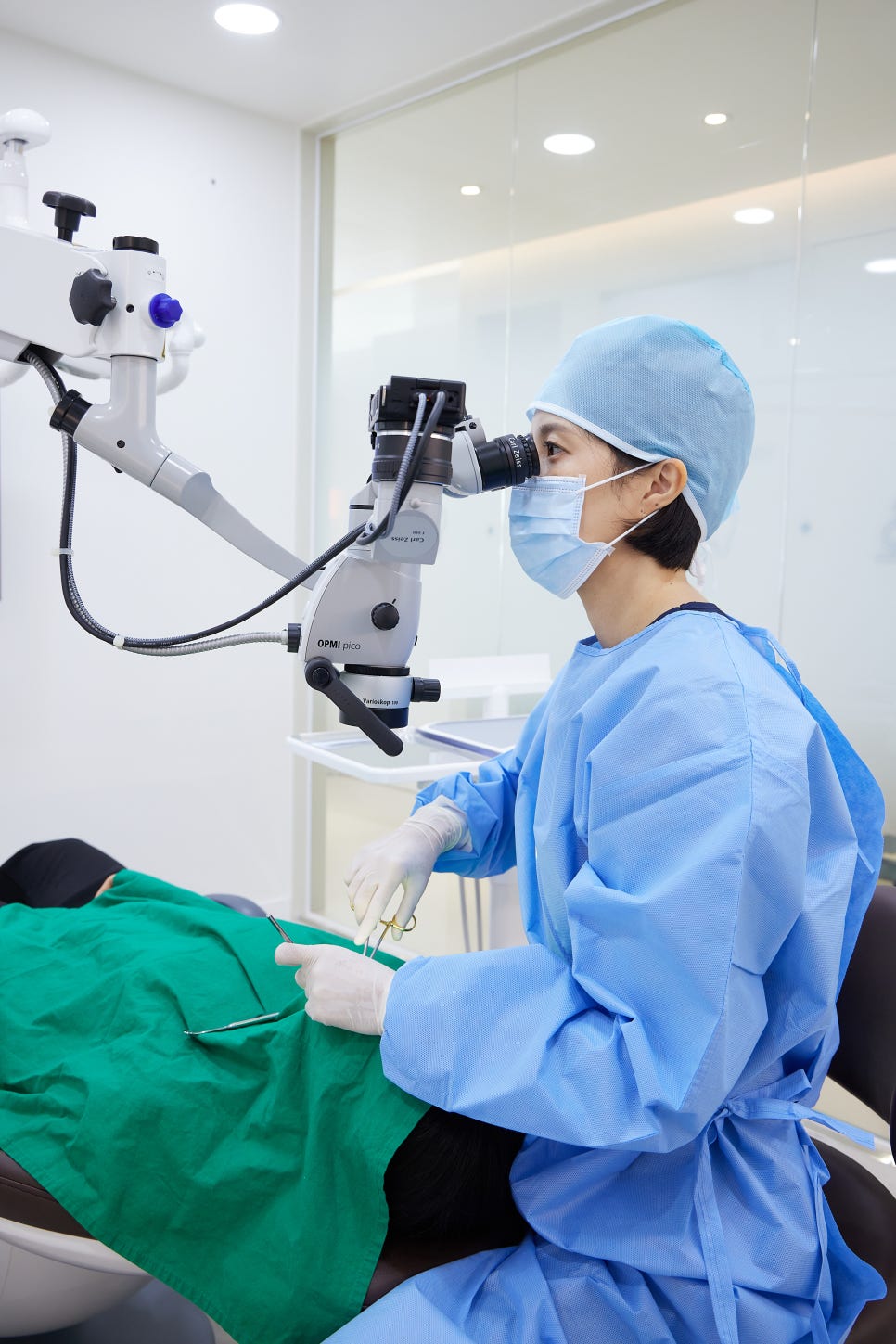
I will explain based on the Carl Zeiss microscope,
a model used at Yonsei University Hospital.
It can provide a field of view magnified up to about 25x,
so when structural limitations or narrowing make it difficult to even detect the canal,
using it allows a wider field of view,
making it easier to find the orifice and disinfect the area.
Even with a 25x magnified view,
the internal structure of the tooth is still very narrow and small, right?! haha
Since there are even more limitations when viewed with the naked eye,
it definitely helps overcome those limitations.
As in the example photo from the dental clinic in Dangsan-dong,
finding the MB2 and DL root allows the NITI file
to enter more smoothly.
Also, because of the presence of pulp stones in the pulp chamber,
it is difficult to identify with the naked eye
whether the root canal orifice has been secured.
Using a microscope allows these issues
to be examined in greater detail.
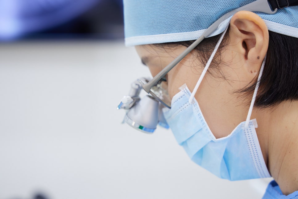
Compared with the loupes commonly used in many hospitals,
a microscope has a much higher magnification! I have shared
why it is a necessary element in treatment.
I hope this post from a dental clinic in Dangsan-dong was helpful
to those who were curious about what kind of equipment it is,
and I will come back next time with another high-quality update.
This has been Kim Min-young,
a specialist in restorative dentistry. Thank you.