Isn’t this just a mole?
When faced with this question,
This morning, I met someone who asked about the brown spots on her face, “This is just a mole, right?”
In fact, I hear this question several times a day in the consultation room.
However, moles and lentigines may look alike on the surface, but the processes that create them within the skin are different.
If you approach them without knowing this difference, it is easy to spend both time and emotional energy without getting the results you hoped for. So today, I would like to point out three things that people considering facial lentigine removal often miss.
Once you have read to the end, you may be able to determine for yourself which one your facial spot more closely resembles.
The person who spent ten years
thinking they were moles
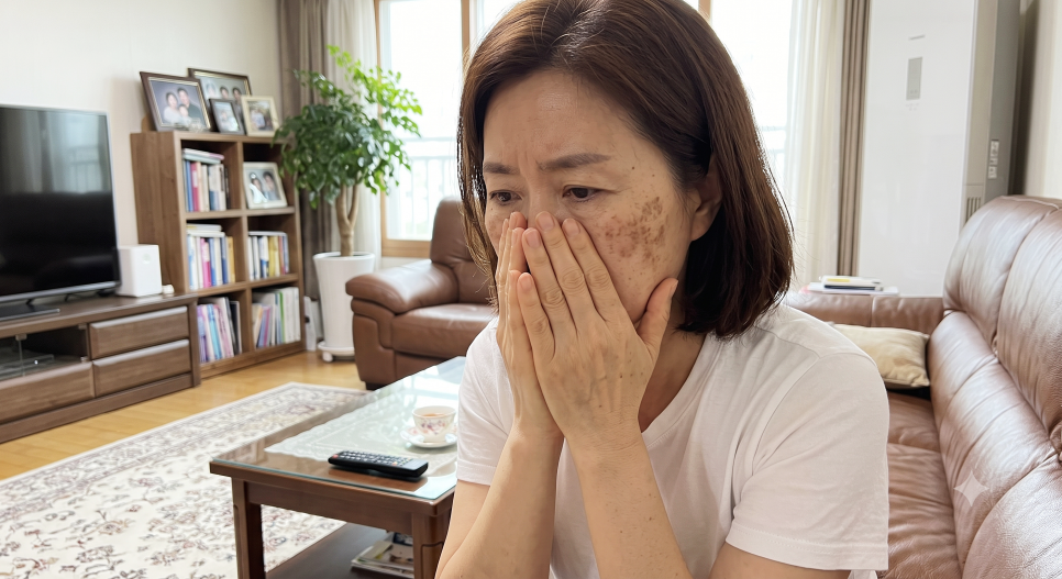
Let me tell you about someone in her late 40s whom I met recently.
She had several light-brown spots
above her cheekbones,
and since she was around twenty,
she had simply assumed
they were moles.
However, over the past few years,
the number had increased,
and the color had gradually
grown darker.
Even when she covered them
with makeup,
they became visible again
by the afternoon,
which made her heart sink
every time she looked in the mirror.
When she searched online, some articles said they could be removed like moles, while others said she would need several laser treatments, which left her even more confused.
The first difference: Where the pigment-producing cells originate
A mole, or nevus, is a condition in which a cluster of cells called nevus cells has gathered within the skin.
I often explain that it is similar to having small grains embedded beneath wallpaper.
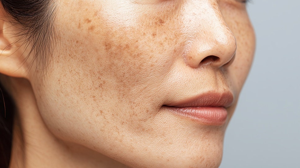
By contrast, a lentigine is a pigmented lesion that develops when melanocytes located in the lowest layer of the epidermis produce more pigment than usual.
Rather than having grains embedded in it, it is more like the wallpaper itself having absorbed color.
That is why, when you touch them with your hand, a mole may sometimes feel slightly raised, while a lentigine is generally flat and level with the surrounding skin.

The second difference: How they respond to sunlight
The second difference is ultraviolet exposure.
Lentigines are known to tend to increase in number or become darker as cumulative sun exposure builds up.
If you feel that they became noticeably darker after the summer, they may be lentigines.
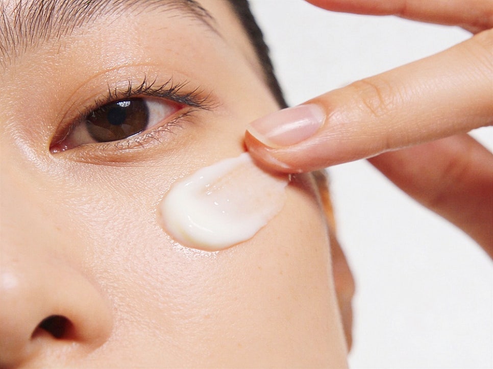
By contrast, a mole that has remained in place for a long time does not change as sensitively in response to sunlight.
This is also why I always discuss sun protection when planning facial lentigine removal.
Leaving the stimuli that darken the pigment unaddressed may lengthen the time needed for management.
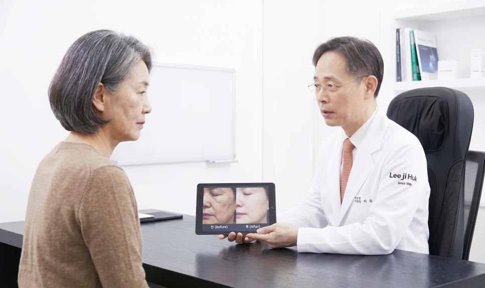
The third difference: The treatment approach is completely different
The third difference is the one most noticeable in the consultation room.
Moles are generally treated by very thinly shaving away the tissue with a CO2 laser.
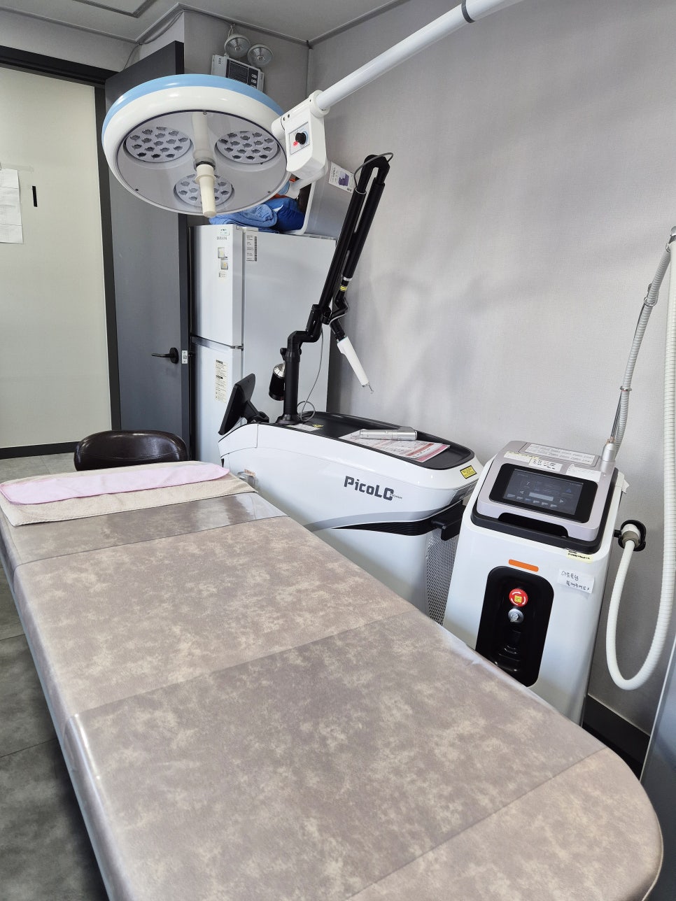
By contrast, for lentigines, devices such as pigment-selective lasers or IPL are used to disperse the color without significantly affecting the epidermis.
In other words, facial lentigine removal is less like digging something out and more like dispersing and lightening the color.
If flat pigmentation is shaved away as though it were a mole, marks may remain. That is precisely why diagnosis should come before treatment.
The answer I gave her
I first suggested that we examine the lesion’s borders and surface with a magnifying glass.
We planned the approach by classifying flat lesions with indistinct borders as more likely to be pigmentation, and raised lesions with distinct borders as more likely to be moles.
Facial lentigine removal is generally carried out over multiple sessions, with intervals between them, rather than being completed all at once while observing the progress.
During the treatment, there may be a stinging sensation like the snap of a rubber band, and numbing cream can be applied if needed.
The color may temporarily appear darker after treatment, or a thin scab may form, but this is often part of the process as it passes.
However, individual differences are possible, so the course may vary.


Rather than dramatic change, a face that remains comfortable over time
She smiled and said that she felt it was a little unfortunate to have spent ten years believing they were moles.
I told her that finding out now was enough.
When it comes to facial lentigine removal, what I always look at first is not how quickly the spots can be erased, but whether the skin at the treated area feels comfortable afterward.
That is also why I have kept the ideas of honest care and proven treatments close to heart for so long.
I hope this post provides a small clue for anyone who has spent a long time hesitating in front of the mirror.

The director answers your questions directly
Q1. How many treatments are usually needed for lentigines?
A. It depends on the depth and extent of the pigmentation.
Because the course differs between pigment located shallowly in the epidermis and pigment located somewhat deeper, treatment is generally divided into multiple sessions at intervals of 2–4 weeks.
Rather than applying a strong treatment all at once, dividing it into sessions at levels the skin can tolerate is known to help reduce the possibility of marks remaining.
Q2. How much should I expect the treatment to cost?
A. The cost varies depending on the number and size of the lesions and the equipment used.
Even among brown spots, the approach differs between those that are more like moles and those that are more like pigmentation, so the cost differs as well.
For that reason, rather than determining a price in advance based only on a photograph or a description, it is less likely to cause misunderstandings if we provide guidance after examining the skin in person.
Q3. Is the recovery process different from mole removal?
A. Yes, it is generally different.
Because tissue is shaved away when removing a mole, a scab often remains for several days, while pigment laser treatment may pass with only slight peeling.
However, sun protection is equally important in both cases.
The recovery pattern may vary from person to person.
Q4. Is there a reason I should have it examined at a dermatology clinic?
A. Moles, lentigines, and seborrheic keratoses are difficult to distinguish based solely on the appearance of a brown spot.
The borders and surface structure need to be examined with a magnifying glass to determine the appropriate direction, and in rare cases, lesions requiring careful attention may also be present.
If the diagnosis is off, the treatment approach will be off as well, so you may feel more at ease by having it checked first.
Q5. Can I receive treatment in the summer?
A. It is possible, but during periods of high ultraviolet exposure, there is a possibility that the pigmentation may become darker again after treatment.
Therefore, in summer, we recommend adjusting the treatment intensity and paying extra attention to sun protection.
If your schedule is flexible, the period from fall to spring offers somewhat more leeway.
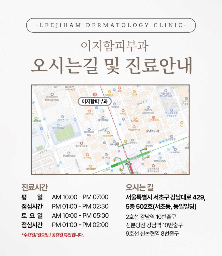
📍 Information about 이지함피부과
🏥 Location
5th floor, Suite 502, 429 Gangnam-daero, Seocho-gu, Seoul (Seocho-dong, Dongil Building)
Near Gangnam Station and Sinnonhyeon Station
☎ Telephone
02-567-5900
🕘 Consultation hours
· Monday, Tuesday, Thursday, Friday 10:00–19:00
· Saturday 10:00–17:00
· Weekday lunch break 13:00–14:30
· Closed Wednesday, Sunday, and public holidays
The clinic is closed on Wednesdays, Sundays, and public holidays, and briefly closes during the weekday lunch break.
Checking the consultation hours before your visit can help reduce your waiting time.